GENOMIC
Mapping
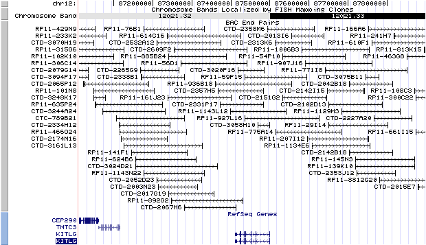
12q21.32. View the map and BAC clones (data from UCSC genome browser).
Structure
(assembly 03/2006)
KITLG isoform a precursor (NM_003994): 9 exons, 87,670 bp, chr12:87410700-87498369.
KITLG isoform b precursor (NM_000899): 10 exons, 87,670 bp, chr12:87410700-87498369.
The figure below shows the structure of the KITLG gene (data from UCSC genome browser).

Regulatory Element
Search the 5'UTR and 1kb upstream regions (seq1=mouse Kitl, seq2=human KITLG) by CONREAL with 80% Position Weight Matrices (PWMs) threshold (view results here).
TRANSCRIPT
RefSeq/ORF
a) KITLG isoform a precursor (NM_003994), 5,351 bp, view ORF and the alignment to genomic.
b) KITLG isoform b precursor (NM_000899), 5,435 bp, view ORF and the alignment to genomic.
Isoform a lacks an alternate in-frame exon, compared to isoform b. Isoform a lacks the primary proteolytic-cleavage site; as a result, the protein encoded by isoform a is largely a membrane bound product. However, isoform b is largely a soluble product.
Expression Pattern
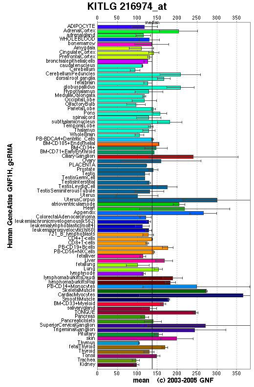
Affymetrix microarray expression pattern in SymAtlas from GNF is shown below.
View more information in Entrez Gene, or UCSC Gene Sorter, or GeneCards.
PROTEIN
Sequence
a) KITLG isoform a precursor (NP_003985): 245 aa, UniProtKB/Swiss-Prot entry Q16493.
b) KITLG isoform b precursor (NP_000890): 273 aa, UniProtKB/Swiss-Prot entry P21583.
Alias: steel factor, stem cell factor, mast cell growth factor.
Ortholog (isoform b)
Species
Chimpanzee Dog Mouse Rat Fowl GeneView
KITLG
KITLG
Kitl
Kitl
LOC396028
Protein
XP_509255 (273 aa)
NP_001012753 (274 aa)
NP_038626 (273 aa)
NP_068615 (273 aa)
NP_990461 (287 aa)
Identities
273/273 (100%) 234/274 (85%) 226/273 (82%) 225/273 (82%) 143/287 (49%)
View multiple sequence alignment (PDF file) by ClustalW and GeneDoc. View evolutionary tree by TreeView.
Domain (isoform b)
(1) Domains predicted by SMART:
Pfam:SCF 1-273
(2) Transmembrane domains predicted by SOSUI:
This amino acid sequence is of a MEMBRANE PROTEIN
which have 2 transmembrane helices.
No. N terminal transmembrane region C terminal type length 1 6 TWILTCIYLQLLLFNPLVKTEGI 28 SECONDARY 23 2 213 LHWAAMALPALFSLIIGFAFGAL 235 PRIMARY 23
(3) Graphic view of InterPro domain structure.
Motif/Site
(1) Predicted results by ScanProsite:
a)N-glycosylation site:
Site : 90 to 93 NISE.
Site : 97 to 100 NYSI.
Site : 118 to 121 NSSK.
Site : 145 to 148 NRSI.
Site : 195 to 198 NDSS.
b) Protein kinase C phosphorylation site:
Site : 54 to 56 TLK.
Site : 119 to 121 SSK.
Site : 126 to 128 SFK.
Site : 192 to 194 SLR.
Site : 200 to 202 SNR.
c) Casein kinase II phosphorylation site:
Site : 80 to 83 SLTD.
Site : 99 to 102 SIID.
Site : 119 to 122 SSKD.
Site : 136 to 139 TPEE.
d) N-myristoylation site:
Site : 27 to 32 GICRNR.
(2) Predicted results of subprograms by PSORT II:
a) Seems to have no N-terminal signal peptide
b) Tentative number of TMS(s) for the threshold 0.5: 2
Number of TMS(s) for threshold 0.5: 1
INTEGRAL Likelihood = -5.31 Transmembrane 216 - 232
PERIPHERAL Likelihood = 4.61 (at 99)
ALOM score: -5.31 (number of TMSs: 1)
c) KDEL ER retention motif in C-terminus: none
d) ER membrane retention signals: none
e) VAC possible vacuolar targeting motif: none
f) Actinin-type actin-binding motif: type 1: none; type 2: none
g) Prenylation motif: none
h) memYQRL transport motif from cell surface to Golgi: none
i) Tyrosines in the tail: too long tail
j) Dileucine motif in the tail: none
(3)Post-translational modifications:
A soluble form is produced by proteolytic processing of isoform 1 in the extracellular domain. Two differentially glycosylated forms are found, LMW-SCF and HMW-SCF. LMW-SCF is fully N-glycosylated at Asn-145, partially N-glycosylated at Asn-90, O-glycosylated at Ser-167, Thr-168 and Thr-180, and not glycosylated at Asn-97 or Asn-118. HMW-SCF is N-glycosylated at Asn-118, Asn-90 and Asn-145, O-glycosylated at Ser-167, Thr-168 and Thr-180, and not glycosylated at Asn-971.
3D Model
(1)
ModBase predicted 3D structure from UCSC Genome Sorter:
Q16493 (KIT ligand isoform a precursor):
none.
P21583 (KIT ligand isoform b precursor):
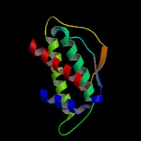
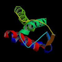
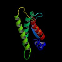
From left to right: Front, Top, and Side views of predicted protein.
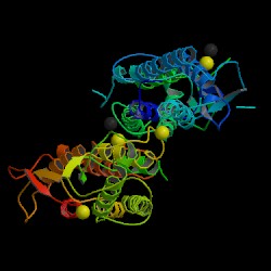
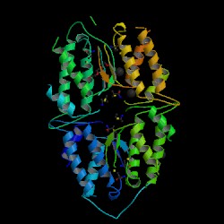
Protein Data Bank (PDB) 3-D Structure of P21583 (KIT ligand isoform b precursor)
2D-PAGE
This protein does not exist in the current release of SWISS-2DPAGE.
Computed theoretical MW=30,898.5Da, pI=5.86 (isoform b).
FUNCTION
Ontology
(1) Biological process: hemopoiesis, epithelial cell proliferation, neural crest cell migration, signal transduction, cell adhesion.
(2) Growth factor activity.
(3) Receptor binding.
(4) Regulation of pigmentation during development.
Location
Plasma membrane, cytoplasm, extracellular space.
Interaction
This gene encodes the ligand of the tyrosine-kinase receptor encoded by the KIT locus. This ligand is a pleiotropic factor that acts in utero in germ cell and neural cell development, and hematopoiesis, all believed to reflect a role in cell migration. In adults, it functions pleiotropically, while mostly noted for its continued requirement in hematopoiesis.
View interactions in HPRD
View co-occured partners in literature searched by PPI Finder.
Pathway
Stimulates the proliferation of mast cells. Able to augment the proliferation of both myeloid and lymphoid hematopoietic progenitors in bone marrow culture. Mediates also cell-cell adhesion. Acts synergistically with other cytokines, probably interleukins.
Cytokine-cytokine receptor interaction , Melanogenesis, Hematopoietic cell lineage in KEGG.
MUTATION
Allele or SNP
No entry for KITLG in HGMD.
SNPs deposited in dbSNP.
Selected allelic examples described in OMIM.
Distribution
In a 6-generation Chinese family with familial progressive hyperpigmentation (FPH), a novel missense mutation c.107A>G (p.N36S) was found (Wang, et al (2009)).
Note that the numbering of the mutations in HGMD is based on KITLG cDNA (NM_000899), view ORF here.
Effect
The N36S missense mutation has a gain-of-function effect on the melanin synthesis (Wang, et al (2009)).
PHENOTYPE
Defect in KITLG causes familial progressive hyperpigmentation (FPH) (Wang, et al (2009)). FPH is an autosomal-dominantly inherited disorder characterized by hyperpigmented patches in the skin, present in early infancy and increasing in size and number with age.
Genetic variations in KITLG are associated with variation in skin/hair/eye pigmentation type 7 (SHEP7) [MIM:611664]. Hair, eye and skin pigmentation are among the most visible examples of human phenotypic variation, with a broad normal range that is subject to substantial geographic stratification. In the case of skin, individuals tend to have lighter pigmentation with increasing distance from the equator. By contrast, the majority of variation in human eye and hair color is found among individuals of European ancestry, with most other human populations fixed for brown eyes and black hair.
REFERENCE
- Wang et al. Gain-of-function mutation of KIT ligand on melanin synthesis causes familial progressive hyperpigmentation. Am J Hum Genet 2009; doi:10.1016/ j.ajhg.2009.03.019.
| Species | Chimpanzee | Dog | Mouse | Rat | Fowl |
| GeneView | KITLG | KITLG | Kitl | Kitl | LOC396028 |
| Protein | XP_509255 (273 aa) | NP_001012753 (274 aa) | NP_038626 (273 aa) | NP_068615 (273 aa) | NP_990461 (287 aa) |
| Identities | 273/273 (100%) | 234/274 (85%) | 226/273 (82%) | 225/273 (82%) | 143/287 (49%) |
View multiple sequence alignment (PDF file) by ClustalW and GeneDoc. View evolutionary tree by TreeView.
Domain (isoform b)
(1) Domains predicted by SMART:
Pfam:SCF 1-273
(2) Transmembrane domains predicted by SOSUI:
This amino acid sequence is of a MEMBRANE PROTEIN
which have 2 transmembrane helices.
No. N terminal transmembrane region C terminal type length 1 6 TWILTCIYLQLLLFNPLVKTEGI 28 SECONDARY 23 2 213 LHWAAMALPALFSLIIGFAFGAL 235 PRIMARY 23
(3) Graphic view of InterPro domain structure.
Motif/Site
(1) Predicted results by ScanProsite:
a)N-glycosylation site:
Site : 90 to 93 NISE.
Site : 97 to 100 NYSI.
Site : 118 to 121 NSSK.
Site : 145 to 148 NRSI.
Site : 195 to 198 NDSS.
b) Protein kinase C phosphorylation site:
Site : 54 to 56 TLK.
Site : 119 to 121 SSK.
Site : 126 to 128 SFK.
Site : 192 to 194 SLR.
Site : 200 to 202 SNR.
c) Casein kinase II phosphorylation site:
Site : 80 to 83 SLTD.
Site : 99 to 102 SIID.
Site : 119 to 122 SSKD.
Site : 136 to 139 TPEE.
d) N-myristoylation site:
Site : 27 to 32 GICRNR.
(2) Predicted results of subprograms by PSORT II:
a) Seems to have no N-terminal signal peptide
b) Tentative number of TMS(s) for the threshold 0.5: 2
Number of TMS(s) for threshold 0.5: 1
INTEGRAL Likelihood = -5.31 Transmembrane 216 - 232
PERIPHERAL Likelihood = 4.61 (at 99)
ALOM score: -5.31 (number of TMSs: 1)
c) KDEL ER retention motif in C-terminus: none
d) ER membrane retention signals: none
e) VAC possible vacuolar targeting motif: none
f) Actinin-type actin-binding motif: type 1: none; type 2: none
g) Prenylation motif: none
h) memYQRL transport motif from cell surface to Golgi: none
i) Tyrosines in the tail: too long tail
j) Dileucine motif in the tail: none
(3)Post-translational modifications:
A soluble form is produced by proteolytic processing of isoform 1 in the extracellular domain. Two differentially glycosylated forms are found, LMW-SCF and HMW-SCF. LMW-SCF is fully N-glycosylated at Asn-145, partially N-glycosylated at Asn-90, O-glycosylated at Ser-167, Thr-168 and Thr-180, and not glycosylated at Asn-97 or Asn-118. HMW-SCF is N-glycosylated at Asn-118, Asn-90 and Asn-145, O-glycosylated at Ser-167, Thr-168 and Thr-180, and not glycosylated at Asn-971.
3D Model
(1)
ModBase predicted 3D structure from UCSC Genome Sorter:
Q16493 (KIT ligand isoform a precursor):
none.
P21583 (KIT ligand isoform b precursor):


From left to right: Front, Top, and Side views of predicted protein.
Protein Data Bank (PDB) 3-D Structure of P21583 (KIT ligand isoform b precursor)


2D-PAGE
This protein does not exist in the current release of SWISS-2DPAGE.
Computed theoretical MW=30,898.5Da, pI=5.86 (isoform b).
(1) Domains predicted by SMART:
Pfam:SCF 1-273
(2) Transmembrane domains predicted by SOSUI:
This amino acid sequence is of a MEMBRANE PROTEIN
which have 2 transmembrane helices.
| No. | N terminal | transmembrane region | C terminal | type | length |
| 1 | 6 | TWILTCIYLQLLLFNPLVKTEGI | 28 | SECONDARY | 23 |
| 2 | 213 | LHWAAMALPALFSLIIGFAFGAL | 235 | PRIMARY | 23 |
(3) Graphic view of InterPro domain structure.
Motif/Site
(1) Predicted results by ScanProsite:
a)N-glycosylation site:
Site : 90 to 93 NISE.
Site : 97 to 100 NYSI.
Site : 118 to 121 NSSK.
Site : 145 to 148 NRSI.
Site : 195 to 198 NDSS.
b) Protein kinase C phosphorylation site:
Site : 54 to 56 TLK.
Site : 119 to 121 SSK.
Site : 126 to 128 SFK.
Site : 192 to 194 SLR.
Site : 200 to 202 SNR.
c) Casein kinase II phosphorylation site:
Site : 80 to 83 SLTD.
Site : 99 to 102 SIID.
Site : 119 to 122 SSKD.
Site : 136 to 139 TPEE.
d) N-myristoylation site:
Site : 27 to 32 GICRNR.
(2) Predicted results of subprograms by PSORT II:
a) Seems to have no N-terminal signal peptide
b) Tentative number of TMS(s) for the threshold 0.5: 2
Number of TMS(s) for threshold 0.5: 1
INTEGRAL Likelihood = -5.31 Transmembrane 216 - 232
PERIPHERAL Likelihood = 4.61 (at 99)
ALOM score: -5.31 (number of TMSs: 1)
c) KDEL ER retention motif in C-terminus: none
d) ER membrane retention signals: none
e) VAC possible vacuolar targeting motif: none
f) Actinin-type actin-binding motif: type 1: none; type 2: none
g) Prenylation motif: none
h) memYQRL transport motif from cell surface to Golgi: none
i) Tyrosines in the tail: too long tail
j) Dileucine motif in the tail: none
(3)Post-translational modifications:
A soluble form is produced by proteolytic processing of isoform 1 in the extracellular domain. Two differentially glycosylated forms are found, LMW-SCF and HMW-SCF. LMW-SCF is fully N-glycosylated at Asn-145, partially N-glycosylated at Asn-90, O-glycosylated at Ser-167, Thr-168 and Thr-180, and not glycosylated at Asn-97 or Asn-118. HMW-SCF is N-glycosylated at Asn-118, Asn-90 and Asn-145, O-glycosylated at Ser-167, Thr-168 and Thr-180, and not glycosylated at Asn-971.
3D Model
(1) ModBase predicted 3D structure from UCSC Genome Sorter:
Q16493 (KIT ligand isoform a precursor):
none.
P21583 (KIT ligand isoform b precursor):


Protein Data Bank (PDB) 3-D Structure of P21583 (KIT ligand isoform b precursor)


2D-PAGE
This protein does not exist in the current release of SWISS-2DPAGE.
Computed theoretical MW=30,898.5Da, pI=5.86 (isoform b).
FUNCTION
Ontology
(1) Biological process: hemopoiesis, epithelial cell proliferation, neural crest cell migration, signal transduction, cell adhesion.
(2) Growth factor activity.
(3) Receptor binding.
(4) Regulation of pigmentation during development.
Location
Plasma membrane, cytoplasm, extracellular space.
Interaction
This gene encodes the ligand of the tyrosine-kinase receptor encoded by the KIT locus. This ligand is a pleiotropic factor that acts in utero in germ cell and neural cell development, and hematopoiesis, all believed to reflect a role in cell migration. In adults, it functions pleiotropically, while mostly noted for its continued requirement in hematopoiesis.
View interactions in HPRD
View co-occured partners in literature searched by PPI Finder.
Pathway
Stimulates the proliferation of mast cells. Able to augment the proliferation of both myeloid and lymphoid hematopoietic progenitors in bone marrow culture. Mediates also cell-cell adhesion. Acts synergistically with other cytokines, probably interleukins.
Cytokine-cytokine receptor interaction , Melanogenesis, Hematopoietic cell lineage in KEGG.
MUTATION
Allele or SNP
No entry for KITLG in HGMD.
SNPs deposited in dbSNP.
Selected allelic examples described in OMIM.
Distribution
In a 6-generation Chinese family with familial progressive hyperpigmentation (FPH), a novel missense mutation c.107A>G (p.N36S) was found (Wang, et al (2009)).
Note that the numbering of the mutations in HGMD is based on KITLG cDNA (NM_000899), view ORF here.
Effect
The N36S missense mutation has a gain-of-function effect on the melanin synthesis (Wang, et al (2009)).
PHENOTYPE
Defect in KITLG causes familial progressive hyperpigmentation (FPH) (Wang, et al (2009)). FPH is an autosomal-dominantly inherited disorder characterized by hyperpigmented patches in the skin, present in early infancy and increasing in size and number with age.
Genetic variations in KITLG are associated with variation in skin/hair/eye pigmentation type 7 (SHEP7) [MIM:611664]. Hair, eye and skin pigmentation are among the most visible examples of human phenotypic variation, with a broad normal range that is subject to substantial geographic stratification. In the case of skin, individuals tend to have lighter pigmentation with increasing distance from the equator. By contrast, the majority of variation in human eye and hair color is found among individuals of European ancestry, with most other human populations fixed for brown eyes and black hair.
REFERENCE
- Wang et al. Gain-of-function mutation of KIT ligand on melanin synthesis causes familial progressive hyperpigmentation. Am J Hum Genet 2009; doi:10.1016/ j.ajhg.2009.03.019.