GENOMIC
Mapping
10q24.32. View the map and BAC contig (data from UCSC genome browser).

Structure
(assembly 07/03)
HPS6/NM_024747: 1 exon, 2,674 bp, chr10:103,489,734 - 103,492,379.
The figure shows the structure of the known isoform (data from UCSC genome browser).

Regulatory Element
Search the 5'UTR and 1kb upstream regions (human and mouse) by CONREAL with 80% Position Weight Matrices (PWMs) threshold (view results here).
TRANSCRIPT
RefSeq/ORF
HPS6/NM_024747: 2,674bp, view ORF and the alignment to genomic.
Expression Pattern
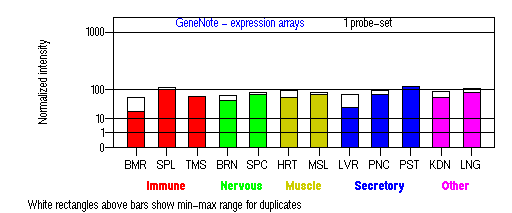
Tissue specificity: ubiquitous, with high expression observed in multiple tissues.

BMR: Bone marrow; SPL: Spleen; TMS: Thymus; BRN: Brain; SPC: Spinal cord; HRT: Heart; MSL: Skeletal muscle; LVR; Liver; PNC: Pancreas; PST: Prostate; KDN: Kidney; LNG: Lung. (data from GeneCards )
PROTEIN
Sequence
HPS6p/NP_079023: 775aa, ExPaSy NiceProt view of Swiss-Prot:Q86YV9.
Synonyms: Hermansky-Pudlak syndrome 6 protein; Ruby-eye protein homolog/Ru.
Ortholog
| Species | Mouse | Rat | Fugu | Zebrafish |
| GeneView | ru/Hps6 | NM_181432 | BK000659 | 13581 |
| Protein | NP_789742 (805aa) | NP_852097 (809aa) | DAA00972 (916aa) | 26509 (680aa) |
| Identities | 82%/635aa | 81%/634aa | 31%/286aa | 32%/230aa |
View multiple sequence alignment (PDF file) by ClustalW and GeneDoc.
Domain
(1) Domains predicted by SMART:
a) signal peptide: 1-20
b) low complexity: 123-139
c) low complexity: 254-268
d) low complexity: 562-577
e) low complexity: 586-604
f) low complexity: 688-708
g) low complexity: 713-730
h) low complexity: 764-772
(2) Transmembrane domains predicted by SOSUI: none.
Motif/Site
(1) Predicted results by ScanProsite:
a) Protein kinase C phosphorylation site [pattern] [Warning: pattern with a high probability of occurrence]:
6 - 8 TlR
196 - 198 ScR, 263 - 265 SpR, 350 - 352 TdR, 398 - 400 SaK, 415 - 417 SlR, 431 - 433 TfR, 521 - 523 TlR, 630 - 632 SgR.
b) Casein kinase II phosphorylation site [pattern] [Warning: pattern with a high probability of occurrence]:
77 - 80 SplD, 263 - 266 SprE, 303 - 306 TlqE, 330 - 333 StlE, 398 - 401 SakD, 422 - 425 TpeE, 617 - 620 TrpE, 755 - 758 SpyE.
c) Cell attachment sequence [pattern] [Warning: pattern with a high probability of occurrence]:
245 - 247 RGD.
(2) Predicted results of subprograms by PSORT II:
a) N-terminal signal peptide: none
b) KDEL ER retention motif in the C-terminus: none
c) ER Membrane Retention Signals: XXRR-like motif in the N-terminus: KRSG
d) VAC possible vacuolar targeting motif: none
e) Actinin-type actin-binding motif: type 1: none; type 2: none
f) Prenylation motif: none
g) memYQRL transport motif from cell surface to Golgi: none
h) Tyrosines in the tail: none
i) Dileucine motif in the tail: none
3D Model
(1) ModBase: none.
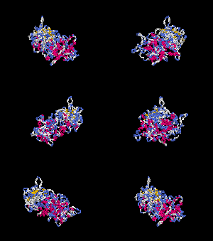
(2) 3D models predicted by SPARKS (fold recognition) below. View the models by PDB2MGIF.


2D-PAGE
This protein does not exist in the current release of SWISS-2DPAGE.
Computed theoretical MW=82,975Da, pI=5.92
FUNCTION
Ontology
Biological process: visual perception.
Location
Cytoplasmic.
Interaction
Zhang, et al reported that HPS6p directly interacts with HPS5p in a complex known as biogenesis of lysosome-related organelles complex-2 (or BLOC2). Gautum, et al found that HPS3p resides in the same protein complex. The native molecular mass of BLOC-2 was estimated to be 340 +/- 64 kDa (view diagram of BLOC-2 complex here). BLOC-2 exists in a soluble pool and associates to membranes as a peripheral membrane protein (Di Pietro, et al).
Pathway
May regulate the synthesis and function of lysosomes and of highly specialized organelles, such as melanosomes and platelet dense granules (view diagram of BLOC-2 pathway here). BLOC-2 functions downstream of BLOC-1 in Tyrp1 trafficking to melanosomes (Setty, et al). BLOC-2, AP-3, and AP-1 coimmunoprecipitated with Rab38 and Rab32 from MNT-1 melanocytic cell extracts,suggesting that BLOC-2, AP-3, and AP-1 proteins function in concert with Rab38 and Rab32 proteins to mediate protein trafficking to lysosome-related organelles (Bultema, et al).
MUTATION
Allele or SNP
SNPs deposited in dbSNP.
1 allelic variant described in OMIM.
Distribution
| Location | Genomic | cDNA | Protein | Type | Ethnicity | Reference |
| Exon 1 | 223C>T | 223C>T | Q75X | nonsense | Italian | Huizing, et al |
| Exon 1 | 238insG | 238insG | D80insG | frameshift, 176X | German | Huizing, et al |
| Exon 1 | 815C>T | 815C>T | T272I | missense | German | Huizing, et al |
| Exon 1 | 913C>T | 913C>T | Q305X | nonsense | Scottish, English, German | Hess, et al; Huizing, et al |
| Exon 1 | 1066ins G | 1066insG | L356insG | frameshift, 366X | Israelian | Schreyer-Shafir, et al |
| Exon 1 | 1234C>T | 1234C>T | Q412X | nonsense | Italian | Huizing, et al |
| Exon 1 | 1712delTCTG | 1712delTCTG | C571delTCTG | frame-shift, 611X | Belgian | Zhang, et al |
| Exon 1 | 1866delTG | 1866delTG | L622delTG | frame-shift, 633X | Irish, German | Hess, et al; Huizing, et al |
| Exon 1 | del 19972bp | del 19972bp | N/A | large deletion | Scottish, English, German | Huizing, et al |
(Numbering of genomic and cDNA sequence is based on the start codon of RefSeq NM_024747.)
Effect
The C571delTCTG mutation resulted in a frameshift with truncation of the nonsense polypeptide at codon 610, causing loss of 30% of the protein at the C terminus (Zhang, et al). Expression analysis revealed no mRNA decay in the fibroblasts of the patient with homozygous 1066C ins G mutation, hence truncated protein is most probably produced (Schreyer-Shafir, et al). Confocal microscopy revealed abnormal distribution of LAMP-3 (lysosomal associated membrane protein-3) in fibroblasts from the HPS6 patient, indicating abnormal trafficking of lysosomal lineage organelles (Schreyer-Shafir, et al). In most HPS6 cases, tyrosinase and TYRP1 are clustered at the perinuclear region of the melanocytes (Huizing, et al).
PHENOTYPE
Defects in HPS6 are the cause of Hermansky-Pudlak syndrome type 6 (HPS-6, OMIM 607522). Zhang, et al described an HPS-6 39-year-old patient with oculocutaneous albinism and easy bruising. She had no pulmonary or gastrointestinal symptoms. Her platelet count was normal, and her bleeding time was moderately prolonged. Platelet function test showed reduced secretion in response to ATP. Electron microscopy of her platelets showed only very rare dense granules. Hess, et al reported that the two HPS-6 patients, similar to HPS-5 patients, exhibited iris transillumination, variable hair and skin pigmentation without the development of pulmonary fibrosis and granulomatous colitis under 27 years old. Clinical studies indicated that HPS-6 patients exhibit oculocutaneous albinism and a bleeding diathesis. Granulomatous colitis and pulmonary fibrosis, debilitating features present in HPS subtypes 1 and 4, were not detected in our HPS-6 patients Huizing, et al.
REFERENCE
- Bultema JJ, Ambrosio AL, Burek CL, Di Pietro SM. BLOC-2, AP-3, and AP-1 proteins function in concert with Rab38 and Rab32 proteins to mediate protein trafficking to lysosome-related organelles. J Biol Chem 2012; 287: 19550-63. PMID: 22511774
- Di Pietro SM, Falcon-Perez JM, Dell'Angelica EC. Characterization of BLOC-2, a complex containing the Hermansky-Pudlak syndrome proteins HPS3, HPS5 and HPS6. Traffic 2004; 5: 276-83. PMID: 15030569
- Gautam R, Chintala S, Li W, Zhang Q, Tan J, Novak EK, Di Pietro SM, Dell'Angelica EC, Swank RT. The Hermansky-Pudlak syndrome 3 (cocoa) protein is a component of the biogenesis of lysosome-related organelles complex-2 (BLOC-2). J Biol Chem 2004; 279: 12935-42. PMID: 14718540
- Hess RA, Claassen DA, White J, Gahl WA, Huizing M. Hermansky-Pudlak syndrome type 5 and type 6: Four new patients. Am J Hum Genet 2003; 73(suppl):456. (Abstract)
- Huizing M, Pederson B, Hess RA, Griffin A, Helip-Wooley A, Westbroek W, Dorward H, O'Brien KJ, Golas G, Tsilou E, White JG, Gahl WA. Clinical and cellular characterisation of Hermansky-Pudlak syndrome type 6. J Med Genet 2009; 46: 803-10.PMID: 19843503
- Schreyer-Shafir N, Huizing M, Anikster Y, Nusinker Z, Bejarano-Achache I, Maftzir G, Resnik L, Helip-Wooley A, Westbroek W, Gradstein L, Rosenmann A, Blumenfeld A. A new genetic isolate with a unique phenotype of syndromic oculocutaneous albinism: clinical, molecular, and cellular characteristics. Hum Mutat, 2006; 27: 1158. PMID: 17041891
- Setty SR, Tenza D, Truschel ST, Chou E, Sviderskaya EV, Theos AC, Lamoreux ML, Di Pietro SM, Starcevic M, Bennett DC, Dell'Angelica EC, Raposo G, Marks MS. BLOC-1 is required for cargo-specific sorting from vacuolar early endosomes toward lysosome-related organelles. Mol Biol Cell 2007; 18: 768-80. PMID: 17182842
- Zhang Q, Zhao B, Li W, Oiso N, Novak EK, Rusiniak ME, Gautam R, Chintala S, O'Brien EP, Zhang Y, Roe BA, Elliott RW, Eicher EM, Liang P, Kratz C, Legius E, Spritz RA, O'Sullivan TN, Copeland NG, Jenkins NA, Swank RT. Ru2 and Ru encode mouse orthologs of the genes mutated in human Hermansky-Pudlak syndrome types 5 and 6. Nat Genet 2003; 33: 145-54. PubMed ID : 12548288
EDIT HISTORY:
Created by Wei Li & Jonathan W. Bourne: 06/18/2004
Updated by Wei Li: 12/25/2006
Updated by Wei Li: 07/28/2012
Updated by Wei Li: 06/13/2013